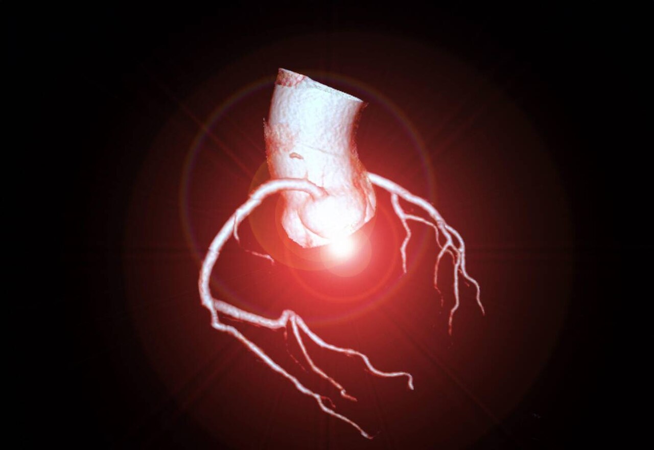
Angiography is an imaging test that shows blood vessels. A special dye, called contrast, is injected into the blood, and X-rays, CT or MRI scans show where it flows. Narrowed, blocked or bulging vessels stand out clearly. It is used to diagnose and treat heart disease, stroke and other vascular conditions.
Why it matters
WHO says cardiovascular diseases are the leading cause of death globally, mostly from heart attacks and strokes [1]. Stroke alone caused 11.9 million new cases in 2021, and 93.8 million people were living after a stroke [2].
Types of angiography
- Coronary angiography: a thin tube (catheter) is passed to the heart arteries, usually through the wrist or groin.
- CT angiography: contrast injected in the arm, with a CT scan; less invasive.
- MR angiography: uses MRI, sometimes without contrast.
- Cerebral angiography: looks at brain vessels, for stroke or aneurysms.
- Peripheral angiography: looks at leg and arm arteries.
From diagnosis to treatment
Catheter angiography is not only a test. During the same procedure, doctors can open blockages with balloons and stents, or remove clots. WHO says some stroke patients may need endovascular thrombectomy, where a clot is pulled out of a brain artery through a catheter [2]. This needs angiography equipment and a trained team, available around the clock.
Risk factors behind the demand
High blood pressure is the leading contributor to stroke, nearly tripling risk [2]. About 1.4 billion adults have hypertension, and only 23% have it under control [3]. Rising rates of diabetes, obesity and ageing populations add to the number of people needing vascular imaging.
Safety
- Contrast dye: can cause allergic reactions or harm kidneys in some patients.
- Radiation: X-ray-based angiography uses radiation; teams aim for the lowest dose possible.
- Catheter risks: bleeding or damage at the entry site, and rare vessel injury.
Using the wrist instead of the groin for coronary procedures has reduced bleeding risks in many centres.
Technology trends
- Lower-dose imaging systems.
- 3D imaging and image fusion to guide complex procedures.
- CT angiography replacing some catheter tests for diagnosis.
- AI tools to measure narrowing and flow.
Access
Over three quarters of cardiovascular deaths occur in low- and middle-income countries [1]. Many of these countries have few cath labs, often only in big cities. Expanding access, with trained staff and reliable supplies, could save many lives.
Key numbers at a glance
- Cardiovascular disease: the leading cause of death.
- 11.9 million new strokes in 2021.
- 1.4 billion adults have hypertension; only 23% controlled.
Go deeper
Want the full picture? Our report covers market size, growth, segments, countries and the key companies. Read the report or get a free sample.
Stroke: time-critical imaging
WHO says that when stroke is suspected, a brain scan should be done as soon as possible [2]. Clot-busting treatment works best within hours [2]. CT angiography shows whether a large brain artery is blocked, which decides whether a patient needs a clot-removal procedure. Stroke networks route these patients to centres with angiography teams on call.
Heart attack care
For a major heart attack, the fastest treatment is to open the blocked artery during angiography. Hospitals measure "door-to-balloon" time, the minutes from arrival to opening the artery. Shorter times mean less heart damage.
Questions for patients
- Why is this test needed, and are there other options?
- Will contrast dye be used, and are my kidneys healthy enough?
- Will the catheter go through the wrist or the groin?
- If a blockage is found, will it be treated during the same procedure?
Peripheral artery disease
Blocked arteries in the legs cause pain when walking and, in severe cases, wounds that do not heal or amputation. People with diabetes and smokers are at higher risk. Angiography shows where blockages are, and balloons or stents can open them. Early diagnosis can save limbs.
Prevention reduces demand
WHO says reducing salt, eating more fruit and vegetables, regular activity and avoiding tobacco and harmful alcohol lower the risk of heart disease and stroke [1]. Every case prevented is one fewer angiogram and procedure needed.
In short
Angiography finds and often fixes blocked vessels in the heart, brain and limbs. Fast access saves heart muscle, brain tissue and limbs.
What this means
Angiography is central to modern heart and stroke care, both for finding problems and treating them. Demand will grow with ageing and chronic disease. Wider access to cath labs outside big cities is the biggest need.
Where to read more
All figures in this article come from public sources listed below. For more detail, start with the WHO cardiovascular disease, stroke and hypertension fact sheets. Each is free to read online. If anything here raises a question about your own health, take this article to your doctor or pharmacist and talk it through together. Buyers, investors and policy makers can also use these sources to check claims in sales materials and compare options on the same public evidence. Where public sources disagree or update their figures, we note the year of each number so readers can see how the picture changes over time. Readers who spot an error or a newer source are welcome to contact our team. Reader questions help us choose which topics to cover in future articles and reports. You can also explore the related market reports, statistics pages and free public health charts linked in the Explore section above, which bring the latest public data on this topic together in one place for easy comparison.
Explore on Acuitas
- Angiography Devices market report – size, growth and key players
- Angiography Devices statistics – key numbers, trials and research in one place
- Top 10 fastest-growing Medical Devices markets
- Global 3D Laparoscopy Imaging Systems Market Analysis – related market
- Global Angioplasty Balloons Market Analysis – related market
- Global Anticoagulants Market Analysis – related market
- Chance of dying 30–70 from heart disease, cancer, diabetes or lung disease – free chart in the Public Health Dashboard
- Clinical trials: Heart and blood vessels – free chart in the Public Health Dashboard
- Get a free sample report or talk to our team
Topics: Cardiovascular · Imaging · Stroke
Sources
Banner image: CT Coronary Artery Imaging by Goleisureintl, CC BY 4.0, via Wikimedia Commons.
The data behind this story
Topics: Cardiovascular · Imaging · Stroke
Go deeper