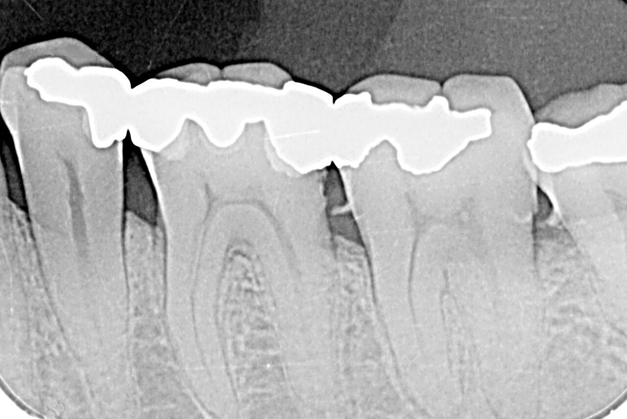
Dentists use X-rays to see what cannot be seen during a normal exam: decay between teeth, bone loss from gum disease, infections at the root, and impacted teeth. Over the past two decades, most practices in richer countries have moved from film to digital imaging. Many now also use 3D scans.
Why imaging matters
WHO estimates that oral diseases affect nearly 3.7 billion people [1]. Untreated tooth decay in adult teeth is the most common health condition worldwide [1]. Severe gum disease affects more than 1 billion cases [1]. X-rays help find these problems early, when treatment is simpler.
Types of dental imaging
- Bitewing X-rays: show the crowns of back teeth to find decay between them.
- Periapical X-rays: show whole teeth and roots.
- Panoramic X-rays: show the whole mouth in one image.
- Cone-beam CT (CBCT): 3D images for implants, complex extractions and root canal work.
- Intraoral scanners: digital impressions without radiation, used for crowns and aligners.
Digital benefits
- Lower radiation dose than film for many exams.
- Instant images, no chemicals.
- Easy to zoom, compare and share with specialists.
- Images can be stored in patient records.
Radiation safety
The American Dental Association says radiation from dental imaging is a minor part of total exposure from all sources [2]. Dentists follow the "ALARA" principle: every X-ray must be justified, doses kept as low as reasonably achievable, and kept well below limits [2]. The FDA and ADA guidance says dentists should order X-rays only when they expect the information to affect patient care [3].
In 2024, the ADA said that modern digital equipment and limiting the X-ray beam to the area being imaged protect patients better. It no longer recommends routine thyroid collars and lead aprons, for all patients including pregnant women [4].
How often?
There is no single schedule. The ADA says how often X-rays are needed depends on a person's oral health, age, risk of disease and symptoms [2]. People at higher risk of decay may need them more often than people with healthy teeth.
Children
The ADA joined more than 80 health organisations in the Image Gently campaign to "child size" X-ray exams for children [2]. Children's doses should be adjusted, and X-rays taken only when needed.
AI in dental imaging
AI tools can now mark possible decay, bone loss and other findings on dental X-rays. They help dentists explain problems to patients and may catch early changes. The dentist still makes the diagnosis.
Key numbers at a glance
- Oral diseases affect nearly 3.7 billion people.
- 23% of adults 60+ have lost all their teeth.
- ADA 2024: thyroid collars and lead aprons no longer routinely recommended.
Go deeper
Want the full picture? Our report covers market size, growth, segments, countries and the key companies. Read the report or get a free sample.
Cone-beam CT
CBCT gives 3D views of teeth, jaws and nerves. It is valuable for planning implants, finding complex root canals and assessing impacted wisdom teeth. It uses more radiation than standard dental X-rays, so it should be used only when 3D information will change treatment. The ADA and the American Academy of Oral and Maxillofacial Radiology published updated selection guidance for 2D and CBCT imaging [2].
Oral cancer
WHO reports that cancers of the lip and mouth had about 389,846 new cases and 188,438 deaths in 2022 [1]. Tobacco, alcohol and areca nut are leading causes [1]. Dental visits, including imaging when needed, are a chance to spot changes early.
Access gaps
In many low- and middle-income countries, dental care and imaging are scarce, especially in rural areas [1]. Portable digital X-ray units and tele-dentistry can extend services, but trained staff and safe equipment are still needed.
Tips for patients
- Tell your dentist if you are pregnant or think you might be.
- Share recent X-rays from other dentists to avoid repeats.
- Ask why an X-ray is needed and how it will change your treatment.
- Keep regular check-ups so problems are found early.
In short
Modern dental imaging is low-dose, fast and precise. Used only when needed, it helps dentists find and treat problems before they become painful or costly.
Records
Keep a copy of your X-rays when changing dentists.
What this means
Digital and 3D imaging have made dental care more precise and safer. With oral disease so common, imaging will keep growing, especially as more countries add dental care to primary health services.
Where to read more
All figures in this article come from public sources listed below. For more detail, start with the WHO oral health fact sheet and the ADA and FDA guidance on dental radiography. Each is free to read online. If anything here raises a question about your own health, take this article to your doctor or pharmacist and talk it through together. Buyers, investors and policy makers can also use these sources to check claims in sales materials and compare options on the same public evidence. Where public sources disagree or update their figures, we note the year of each number so readers can see how the picture changes over time. Readers who spot an error or a newer source are welcome to contact our team. Reader questions help us choose which topics to cover in future articles and reports. You can also explore the related market reports, statistics pages and free public health charts linked in the Explore section above, which bring the latest public data on this topic together in one place for easy comparison.
Explore on Acuitas
- Dental X-Ray Equipments market report – size, growth and key players
- Dental X-Ray Equipments statistics – key numbers, trials and research in one place
- Top 10 fastest-growing Medical Devices markets
- Global 3D Laparoscopy Imaging Systems Market Analysis – related market
- Global Angiography Devices Market Analysis – related market
- Global Artificial Intelligence (AI) in Medical Imaging Market Analysis – related market
- MRI scanners – free chart in the Public Health Dashboard
- CT scanners – free chart in the Public Health Dashboard
- Get a free sample report or talk to our team
Sources
- WHO fact sheet: Oral health
- American Dental Association: X-rays/radiographs
- US FDA: The selection of patients for dental radiographic examinations
- ADA News (Feb 2024): Using thyroid collars during radiographic exams no longer recommended
Banner image: X-ray of dental calculus by Jmarchn, CC0, via Wikimedia Commons.
The data behind this story
Go deeper