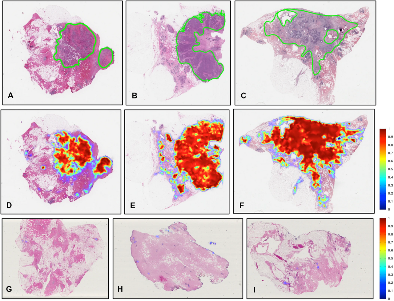
Pathologists diagnose disease by looking at thin slices of tissue on glass slides under a microscope. It is central to cancer diagnosis. Digital pathology scans those slides into very high-resolution images that can be viewed on a screen, shared instantly and analysed by software.
A regulatory milestone
In April 2017, the US FDA allowed marketing of the first whole slide imaging system for reviewing and interpreting digital surgical pathology slides made from biopsied tissue [1]. This opened the door for pathologists in the US to make primary diagnoses from screens, not just microscopes.
Why demand is growing
Cancer cases are rising. The WHO cancer agency estimated 20 million new cancer cases in 2022, and expects over 35 million in 2050 [2]. Nearly every suspected cancer needs a pathology diagnosis.
At the same time, many countries have too few pathologists. The Lancet Commission on diagnostics highlighted major shortages of diagnostic staff, especially in low- and middle-income countries [3]. Digital slides can be read by a pathologist anywhere, which helps spread scarce expertise.
How it works
- Tissue is processed and placed on glass slides, as usual.
- A slide scanner captures the whole slide at high magnification.
- Images are stored in a secure system linked to the lab record.
- Pathologists view, zoom and measure on a high-quality screen.
- Software tools can count cells, measure areas or flag regions of concern.
Benefits
- Remote reading: second opinions and expert reviews without posting slides.
- Workload sharing: spreading cases across a network of labs.
- Teaching: sharing rare cases with trainees anywhere.
- Measurement: more consistent counts and scores.
- AI: digital images make computer-aided analysis possible.
Challenges
- Cost: scanners, storage and screens are expensive.
- Data size: one slide can be several gigabytes; a lab may make thousands a day.
- Workflow: scanning adds a step and needs reliable, fast systems.
- Validation: each lab must show digital reads match glass reads.
- Payment: few systems pay extra for digital or AI reads.
AI in pathology
AI tools can help find cancer in biopsies, grade tumours and predict gene changes from images. Some have regulatory clearance for specific uses. Like AI in radiology, they work best as helpers that save time and add a second look, with the pathologist making the final call.
Key numbers at a glance
- First FDA-authorised whole slide imaging system for primary diagnosis: April 2017.
- New cancer cases: 20 million (2022), over 35 million expected by 2050.
Questions for labs going digital
- Which cases will we scan first?
- How will we store and back up images?
- How will we validate digital against glass?
- Which AI tools are cleared for our uses?
Go deeper
Want the full picture? Our report covers market size, growth, segments, countries and the key companies. Read the report or get a free sample.
Cancer burden drives pathology demand
WHO's cancer agency reported 9.7 million cancer deaths in 2022 [2]. Lung, breast and colorectal cancers were the most common [2]. Each diagnosis, and often each stage of treatment, depends on pathology. Tumour gene testing, which guides targeted drugs, adds more work for labs.
Telepathology for remote areas
In many low- and middle-income countries, there are very few pathologists outside big cities. Digital slides can be scanned at a district hospital and read by an expert far away. This can cut the time to diagnosis from weeks to days. Reliable internet and trained staff to prepare good slides are essential.
Quality starts with the slide
A scanner cannot fix a poor slide. Tissue must be handled, cut and stained well. Labs going digital often find they need to improve these earlier steps. Good slides improve both human and AI reads.
Steps for a lab going digital
- Start with a clear goal, such as faster second opinions or remote reading.
- Scan a pilot set of cases and compare digital with glass reads.
- Plan storage, backup and network speed before scaling up.
- Train pathologists and technicians on the new workflow.
- Add AI tools only after the digital workflow runs smoothly.
Labs that phase the change in steps tend to avoid disruption and build confidence among staff. Partnerships between labs can share the cost of scanners and storage.
Who benefits most
Patients in remote areas gain faster diagnoses. Pathologists gain flexible working and easier second opinions. Hospitals gain the ability to share workload across sites. Researchers gain large image libraries for study and AI training, with proper consent.
What this means
Digital pathology is moving from early adopters to mainstream labs. It helps share scarce pathologists, supports AI and speeds second opinions. Costs and data storage are the main hurdles, but rising cancer numbers make the case stronger each year.
Where to read more
All figures in this article come from public sources listed below. For more detail, start with the FDA's 2017 notice on whole slide imaging and the IARC global cancer estimates. Each is free to read online. If anything here raises a question about your own health, take this article to your doctor or pharmacist and talk it through together. Buyers, investors and policy makers can also use these sources to check claims made in sales materials, and to compare products and programmes on the same public evidence. Asking for the original source behind any number is a simple habit that leads to better decisions.
Explore on Acuitas
- Digital Pathology Systems market report – size, growth and key players
- Digital Pathology Systems statistics – key numbers, trials and research in one place
- Top 10 fastest-growing Digital Health markets
- Global Alzheimers Dignostic Market Analysis – related market
- Global Antimicrobial Susceptibility Testing Market Analysis – related market
- Global Artificial Intelligence (AI) in Drugs Discovery Market Analysis – related market
- Early deaths from long-term diseases (WHO) – free chart in the Public Health Dashboard
- Clinical trials: Cancer – free chart in the Public Health Dashboard
- Get a free sample report or talk to our team
Topics: Diagnostics and lab testing · Oncology · Artificial intelligence
Sources
- US FDA (Apr 2017): FDA allows marketing of first whole slide imaging system for digital pathology
- WHO/IARC news (1 Feb 2024): Global cancer burden growing
- The Lancet Commission on diagnostics (2021)
Banner image: Histopathology of tumor identification by humans and AI by Cruz-Roa A, Gilmore H, Basavanhally A, Feldman M, Ganesan S…, CC BY 4.0, via Wikimedia Commons.
The data behind this story
Topics: Diagnostics and lab testing · Oncology · Artificial intelligence
Go deeper